


o Visual field: the perceptual space available to the fixating eye
o Purpose: to provide a gross check for any defects in the peripheral visual field
O Extinction phenomenon
І Patients with right parietal lesions can exhibit a form of visual extinction. When shown two objects, one contralateral (left) and one ipsilateral (right) to the lesioned hemisphere, subject will report seeing only the one in the ipsilateral (right) field
o RiddochТs phenomenon
І Some patients with neurological defects suffer from stato-kinetic dissociation
Ј Moving objects are perceived better than static ones
Ј Defects present on automated perimetry (static) tend to be more extensive compared to those measured by manual perimetry (kinetic)
O Finger counting
І Tests the patientТs ability to correctly identify gross targets in each of the 4 major quadrants
І Procedure
Ј Examiner and patient remove spectacles
Ј Sit at eye level and 1m away
Ј Have patient occlude OS with palm of their hand and fixates clinicianТs OS with their OD (clinicianТs visual field corresponds to the patientТs)
Ј Place one hand in the mid-plane (50 cm) at about 45∞ from fixation
o Important to be exactly between you and the patient so the patientТs field can be compared to yours
І Fingers more than 50 cm from patientà patientТs field will be underestimated/constricted
І Fingers are less than 50 cm from patientà field will appear to be normal but you may be more likely to miss a defect/constriction
Ј Present one, two, or four fingers in one of the four quadrants
Ј Repeat for other 3 quadrants
Ј Present both hands simultaneously in both superior quadrants
Ј Present the fingers of both hands and ask patient to add together
o Do NOT use the same numbers in each hand
Ј Repeat for OS
Ј Record normal fields as FTFC (full to finger counting) OD, OS
o If not full, then document/draw constricted quadrant
І Advantages
Ј Sensitive to homonymous (neurologic) quadrantic and hemianopic VF defects
Ј Fast and can be performed in any location
Ј Can test for extinction phenomenon
І Disadvantages
Ј Results are not meaningful to the DMV
Ј Sensitivity is not very high
Ј Limits of the VF are not tested
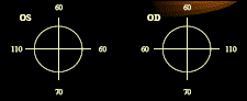
O Field Limits
І Compares known peripheral field limits to the patientТs peripheral field limits
І Procedure
Ј Patient removes spectacles and occludes OS; have patient fixate your nose
Ј Move target (wand) from behind patient (non-seeing to seeing) toward the horizontal limit of the field
o Test slightly above and below the temporal midline
Ј Have patient tell you when it comes into view
Ј Do the same for the superior and inferior visual field
o Test on both sides of the superior and inferior midline
Ј Test nasal side
o Test on either side of the nasal midline
Ј Repeat for OS
Ј Record limits (ALWAYS record from the patientТs perspective)
o Normal
І Advantages
Ј Provides a means to quantify confrontation fields
|
|
|
Ј Easier for patient to understand and/or respond
І Disadvantages
Ј Testing the limits of the VF produces variable sensitivity, therefore difficult to detect true visual field loss in the far peripheral field
Ј Does not screen for extinction phenomenon
V Interpupillary Distance
o Distance between centers of the entrance pupils
І Important for:
Ј Alignment of optical instruments (avoids prismatic effects induced)
Ј Spectacle design considerations
o Optical centers match PDТs (if not, induces prism)
Ј Documentation of craniofacial abnormalities
І Measure monocular PDТs for high powered spectacle prescriptions, PALs
o Procedure for binocular PD
І Sit at eye level with patient ~40 cm away
І Close your right eye and have patient look into your open left eye
І Place zero at the temporal limbus of the right eye (DO NOT MOVE)
І Note position that is aligned with the nasal limbus of the left eye: NEAR PD
І Close left eye and have patient look into your open right eye
І Note position that is aligned with the nasal limbus of the right eye: DISTANCE PD
І Record distance/near
o Procedure for monocular PD
І Place ruler on the patientТs bridge
І Close your right eye and have patient look into your left eye
І Align zero mark with the center of the pupil (CANNOT use pupil margin or limbus)
І Note the mark centered on the bridge: OD MONOCULAR PD
І Move ruler and place an easily recognized mark on the center of the bridge (use this as the zero mark)
І Open your right eye, close your left, and have patient look into your right eye
І Note mark centered in the patientТs left pupil; subtract the СzeroТ reading from the last reading: OS MONOCULAR PD
o Use PrenticeТs rule to calculate the induced prism from decentration
Ј P= dF
o Errors: unsteady positioning, error in parallax, patientТs with fixation disparities and doctorТs PD significantly wide (will overestimate)
V Ocular Dominance
o The preferential sighting of a target with one eye
o In monovision CLs fitting, the dominant eye is generally fit with the distance
o Useful when the subjective match in the clarity of the lines of letters cannot be achieved during binocular balance
І Leave dominant with slightly clearer vision
o Do not leave the VA of the dominant eye worse than the non-dominant eye
o Place prism before the non-dominant eye
o Procedure
І Instruct patient to fully extend arms and create a triangle with both hands
І Patient looks through aperture at the doctorТs right eye
Ј Eye aligned with the doctorТs is the dominant eye
І Record ocular dominance

