


1. Give difinition of the Histology.
2. Give difinition of the Cytology.
3. Give difinition of the Embryology.
4. Give difinition of the Cell.
5. The structure of the Plasmolemma.
6. Characteristic functions of the plasmolemma:
a) receptive function;
b) transport function.
7. The structure of intercellular contacts:
- Simple contact;
- Zonular occludentes;
- Synapse;
- Desmosome;
- Zonular adherents;
- Gap junctions.
8. Characteristic and structure of symplast and sincytium.
Theme: The structure of the Cytoplasm.
1. The structure of the Cytoplasm.
2. The structure and functions of Mitochondria.
3. The structure and functions of Lysosomes and Peroxisomes.
4. The structure and functions of Peroxisomes.
5. The structure and functions of Agranular Endoplasmic Reticulum.
6. The structure and functions of Granular Endoplasmic Reticulum.
7. The structure and functions of Ribosomes.
8. The structure and functions of Golgi Bodies.
Theme: The structure of the Cytoplasm.
1. The structure and functions of Microfilaments.
2. The structure and functions of Microtubules.
3. The structure and functions of Cytocentrum (centrosome).
4. The structure and functions of Cilia and Flagella.
5. Name of types Inclusions and their functions.
Practical pat.

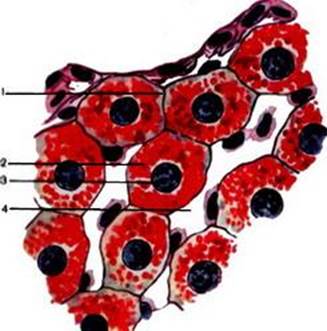
Slide є1 Liver cell (hepatocytes).
Staining: hematoxylin Ц eosin.
1 Ц hepatocyte; 2 Ц nucleus; 3 Ц cytoplasm; 4 Ц cell membrane; 5 Ц capillaries.

 1
1
 2
2
Slide є2 Symplast.
Staining: iron hematoxylin.
1 Ц nucleus; 2 Ц transverse-striated.

Slide є3 Intercellular substance (elastic cartilage).
Staining: orsein.
1 Ц basal substance; 2 Ц network of elastic fibres; 3 - chondrocytes.

Slide є4 Mitochondria.
Staining: hematoxylin Ц eosin.
1 Ц mitochondria; 2 Ц nucleus; 3 Ц lumen of the tubules kidney.

 3
3
Slide є5 Golgi apparatus in the nerve cell of spinal ganglia.
Staining: osmium Ц impregnated.
1 Ц nucleus; 2 Цcytoplasm; 3 Ц Golgi apparatus.
Slide є6 Glycogen inclusions in the liver cells (hepatocytes).
Staining: BestТs method.
1 Ц hepatocyte; 2 Ц glycogen granules; 3 Ц nucleus; 4 - cappillaries.

Slide є7 Lipid inclusions in the liver cells.
Staining: safranin and osmium Ц impregnated.
1 Ц hepatocyte; a Ц lipid inclusions; b Ц nucleus; 2 - capillaries.
 2 1
2 1

Slide є8 Pigment inclusions in the melanocytes.
Staining: slide is not stained.
1 Ц nucleus; 2 Ц pigment granules in the cytoplasm; 3 Ц cell processes.


 6 1a
6 1a
 |
 1b
1b
 1
1
Diagram є1 Structure of the plasmolemma.
1 Ц lipid molecule: a) head; b) tail; 2 Ц elementary biological membrane; 3 Ц half integral proteins; 4 Ц integral proteins; 5 Ц peripheral proteins; 6 Ц glycocalyx; 7 Ц cortical layer; 8 Ц microfilaments; 9 Ц microtubules; 10 Ц glycoproteinТs; 11 Ц glycolipids.
|
|
|

Diagram є2 Zonular occludentes.
1 Ц plasmolemma; 2 - integral proteins; 3 Ц microfilaments.

Diagram є3 Desmosomes.
1 Ц plasmolemma; 4 Ц tonofilaments.

Diagram є4 Gap junctions (nexus).
1 Ц plasmolemma; 2 - integral proteins; 5 Ц connexons.
LECTURE є 2
Topic: THE NUCLEUS OF THE CELL. REPRODUCTION OF CELLS. AGING AND DEATH OF CELLS.
NUCLEUS is the most important component of the cell, containing its genetic apparatus.
The nucleus functions:
1. Storage of genetic information (containing DNA constant structure).
2. Reproduction and transmission of genetic information (duplication of genetic material and division between daughter cells).
3. Realization of genetic information (formation protein synthesis apparatus).

