


Excitability changings
(figure of action potentials phases and excitability changings correlation)
| Action potential phase | Excitability changing | Reasons and mechanisms |
| Partial depolarization | Supernormal period | The less threshold is, the more excitability is |
| Complete depolarization | Absolute refractiveness (non-excitability) | During overshoot cellular excitability is equal to zero due to Na-channels inactivation and K-channels activaton. Membranes can not react even to epiliminal stimuli. Potentials difference is equal to 0. |
| Rapid repolarization | Relative refractive-ness | K-ions come from cell and negative charge is accumulated on internal membrane surface. Excitability is restored. Membrane can react to superliminal stimuli. Substances prolonging relative refractory period (antiarhythmical) decrease cardiac contraction rate and repair heart rhythm. |
| Slow repolarization | Supernormal period (exaltation) | Membrane is partially depolarized and is excited very easy. Answer reaction can occur even at subliminal stimuli action. |
| Hyperpolari-zation | Subnormal excitability | Membrane potential increasing, threshold increasing define excitability decreasing. Besides, hypoexcitability is delt with Na-channels inactivation and K-ions activation. Only epiliminal stimuli can cause answer reaction. |
3. To designate graphically muscles contraction types dependently on irritation rate.
At a muscle irritation by single stimulus the single muscular contraction arises. One can distinguish the latent period lasting 0,01 sec (from irritation beginning to answer-back reaction beginning), shortnening period (actually contraction) lasting 0,04 sec and relaxation period lasting 0,05 sec. Thus, singular muscular contraction lasts 0,1 sec.
In reply to a rhythmic irritation (namely the such one our muscles are received) the muscle is reduced lengthly (for a long time). Such contraction has received the name tetanic or summarized. If each subsequent pulse approaches to a muscle in the period, when it began to be relaxed, there is an infused, dentate or incomplete tetanus.
If the interval between irritations decreases so, that each subsequent pulse comes to a muscle, at that moment, when it is in a contraction phase, there is smooth, complete, fused tetanus or holotetanus.
4. Electromyography Ц the method principle, its types and main indexes. Masticatory muscles electromyography Ц for dental faculty students.
EMG - With other words, it is registration method of skeletal muscles excitability by electrical potential oscillations occurrence under rest, at tonic tension and arbitrary movements. There are 3 main electromyogram kinds (by square of record and electrodes:
Ј interferential Ц muscular biopotentials are taken off from large surface while applying the electrodes on skin;
Ј local Ц separate motor units activity is registered by means of needle electrodes;
Ј stimulatory Ц the registration of electrical muscle answer to the stimulation of nerve innervating it.
|
|
|
On other hand (by appearance):
a) saturated - in norm Ц impossible to be analyzed;
b) non-saturated Ц possible to be analyzed quantitatively Ц oscillations are more seldom.
2 analysis types:
1) qualitative Ц the curve description;
2) quantitative Ц to estimate rate and amplitude.
Main branches of application:
1) neurology.
2) traumatology-orthopedy,
3) pediatry,
4) sportive medicine and rehabilitation medicine,
5) dentistry all branches.
5. To draw reflectory arc, to designate its main elements.
In AhmedТs copy-book
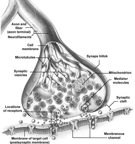
6. To draw synapse, to explain excitement conductance mechanism.
| Presy≠naptic neuron |  Arrive of action potential in axon terminal Arrive of action potential in axon terminal
|
 Opening of calcium channels in presynaptic membrane Opening of calcium channels in presynaptic membrane
| |
 Influx of calcium ions from ECF into the axon terminal Influx of calcium ions from ECF into the axon terminal
| |
 Opening of vesicles and release of Ach Opening of vesicles and release of Ach
| |
| Passage of Ach through synaptic cleft | |
| Postsy≠naptic neuron |  Formation of Ach-receptor complex Formation of Ach-receptor complex
|
 Development of EPSP Development of EPSP
| |
 Opening of sodium channels and influx of sodium ions from ECF Opening of sodium channels and influx of sodium ions from ECF
| |
 Opening of sodium channels in initial segment of axon Opening of sodium channels in initial segment of axon
| |
| Influx of sodium ions from ECF and development of action potential
| |
| Spread of action potential through axon of postsynaptic neuron |
Fig.15. Sequence of events during synaptic transmission. Ach = Acetylcholine. ECF = Extracellular fluid. EPSP = Excitatory postsynaptic potential
PROPERTIES OF SYNAPSE
1. ONE WAY CONDUCTION (BELL-MAGENDIE LAW)
According to Bell-Magendie law, impulses are transmitted only in one direction in synapse, i.e. from presynaptic neuron to postsynaptic neuron.
 2. SYNAPTIC DELAY
During transmission of impulses via the synapse, there is a little delay in the transmission. This is called synaptic delay. This is due to the time taken for:
a) Release of neurotransmitter
b) Movement of neurotransmitter from axon terminal to postsynaptic membrane for mediator interaction with receptor
c) Action of the neurotransmitter to open the ionic channels in postsynaptic membrane
d) Mediator restoration.
The synaptic delay is one of the causes for the latent period of the reflex activity.
3. FATIGUE
The fatigue at the synapse is due to neuro-tansmitter substance, acetylcholine, level decreasing and mediator exhausting. After producing the action, this neurotransmitter is destroyed by acetylcholinesterase. Synapse is the mostly-fatigable structure in nervous system (compare: nervous fiber is practically non-fatigable).
4. SUMMATION
When many presynaptic excitatory terminals are stimulated simultaneously or when single presynaptic terminal is stimulated repeatedly, there is summation in postsynaptic neuron. This is called summation. Summation is of two types.
1) Spatial Summation
This occurs when many presynaptic terminals are stimulated simultaneously (for example, in central synapses).
2) Temporal Summation
It occurs when one presynaptic terminal is stimulated repeatedly. Also subliminal stimuli summation (3-5) to form EPSP can be proper example.
Thus, both spatial summation and temporal summation play an important role in the facilitation of response.
5. ELECTRICAL PROPERTIES
The electrical properties of the synapse are the EPSP and IPSP.
6. DECREASING TRANSFORMATION OF RHYTHM INTO FREQUENCY. 3-5 subliminal stimuli on entrance will give 1 action potential on exit.
|
|
|
7. To draw spine reflexes reflectory arcs, to call closage level.
Abdominal reflexes:
a)superior Ц itТs caused by puncture irritation of abdomen skin in parallel of rib arc; the reflexes arc is closed at D6-D8 segments of spinal cord (from D Ц dorsal);
b) intermediate Ц by similar irritation but at horizontal dimension at navel level; the reflexes arc is closed at D9-D10;
c) inferior- in parallel to groin plica; the reflexes arc is closed at D11-D12.
Plantar reflex - is a plantar flexion of foot toes as a response of puncture irritation of external plantar limb; the reflexes arc is closed at L5-S2 and is in sciatic nerve.
At injury of corresponding motor nerve and corresponding link of reflector arc the response reaction is decreased or disappeared (areflexy), muscular atony, atrophy are observed.
Biceps-reflex Ц is caused by irritation of muscle tendon above cubital joint by neurologic hammer. Answer reaction Ц hand flexion in cubital joint. The reflexes arc is closed at C5-C6. Afferent and efferent fibers are in muscular-cutaneous (skin) nerve structure.
Triceps-reflex - is caused by hammer shock on triceps-muscle tendon, on 1-1,5 cm upper of posterior process of ulna. Answer reaction- muscular contraction and antebrachium (fore-arm) extension. The reflexes arc is closed at C6-C8. The fibers are in medianus, radialis and muscular-cutaneous nerves.
Carpo-radialis reflex Цis investigated by hummer shock onto awl-like processus of radius. Answer reaction Ц flexure in cubital joint and antebrachium pronation. Origin location: investigated person hand must be bended at obtuse angle in cubital joint; personТs examinated hand is supported by doctorТs hand at the locus between pronation and supination. The reflexes arc is closed at C5-C8. The fibers are in medianus, radialis and muscular-cutaneous nerves.
Knee jerk or patellar tendon reflex - is caused by light shock of hummer on musculus quadriceps femoris tendon. Answer reaction Ц tibia extension. The reflexes arc is closed at L2-L4. Sensor and motor fibers are in femoral nerve.
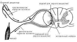
„увствительное волокно Ц sensory (afferent) fiber.
ƒвигательное волокно Ц motor fiber
«адний рог серого вещества Ц grey substance posterior horn (corn)
ѕередний рог Ц anterior corn (horn)
¬ставочный нейрон - associative neuron
ћышца Ц muscle
8. To draw pyramidal tract, to designate peripheral and central neurons.

Central or the 1st neuron Ц pre-central gyrus, giant pyramids of Betz.
Peripheral neuron or the 2nd Ц spine anterior corn.
9. To explain symptoms at pyramidal tract central and peripheral neuron damage.
| Paralysis type | Central or spastic | Peripheral, sluggish or atrophic |
| Injuries location | Cortex motor projectional area or pyramidal fascicles | Spine anterior corn, peripheral nerves anterior fascicles and anterior fibers |
| Paralysis distribution | More often diffused | More often limited |
| Muscles tone | Hypertony, spasticity | Hypotony, atrophy |
| Reflexes | Deep reflexes are increased, abdominal and plantar are decreased or lost | Deep and skin reflexes are decreased or lost |
| Pathological reflexes | Some of them are present | Absent |
| Conjugating movement | Present | Absent |
| Degeneration reaction | Absent | Present |
10. To draw superficial sensitivity conductive pathways.

Fig.22. Spinothalamic tracts and pathways for crude touch, pain and temperature sensations. Anterior spinothalamic tract carries crude touch sensation. Lateral spinothalamic tract carries pain and temperature sensations.
The 1st neuron Ц spinal ganglion.
The 2nd neuron Ц thalamus.
The 3rd neuron Ц cortex post-central gyrus.
11. To draw prophound (deep) sensitivity conductive pathways.
Ways of Goll and Burdah.

Gracilis Ц of Goll, cuneatus or cuneate Ц of Burdah.
|
|
|
12. To call and to show reflexes of posterior brain that allows assessing cranial-cerebral nerves functions.
Corneal reflex Ц the investigated person looks up and towards. The investigator touches with thin paper strip to the inferior-exterior eyeside without touching the eyelashes. The reflex arc - orbital nerve (Vth pair ramus), pons, facial nerve. Decreasing or lost of corneal reflex is found out at trigeminal nerve, facial nerve, pons injuries, at shock, in course of narcosis.
Conjunctival reflex Ц is caused by touching to conjunctive. Answer reaction- eyelid close. Reflectory arc Ц see like at corneal reflex.
Superciliar reflex - is caused by hummer shock at superciliar arc limb. Answer reaction - eyelid closure. Reflex arc Ц orbital nerve, pons, facial nerve.
Mandibular reflex - the investigated person slightly opens his mouth. Masticatory muscles contraction is caused by hammer shock down on chip from one than from another side. Answer reaction Ц mandible lifting. This reflex can be absent under normal conditions.
13. What is the difference between static and static-kinetic reflexes of midbrain?
Static reflexes - tonic reflexes under resting conditions (while standing, sitting, head turns and inclinations, as well as arms movements, legs movements while sitting. Role: natural posture saving under rest.
Statho-kinetic reflexes Ц are realized at posture changings during movements and include:
Ј head straitening (from vestibular and skin receptors),
Ј trunk straitening (from skin proprioreceptors and skin receptors),
Ј Straitening, establishment reflexes (tone redistribution at posture changing).
Role: natural posture maintaining at movements.
14. Cerebellum. Probes of its functions assessment and injury main symptoms.
Signs of a cerebellum lesion
Static ataxy Ц it is infringement of statics in rest (shaking).
Dynamic ataxy Ц it is locomotion coordination disorder at performance of targeted movements. A tremor of extremities Ц so-called intentional tremor (during movements, stops during rest) Ц is observed.
Walking is shaky, УdrunkФ.
Cause of cerebellar ataxy is the disturbance of сonjugative work of muscles Ц agonists and antagonists, disproportion of locomotion Ц dysmetria. Rapid change of one locomotion by others, inverse ones Ц adiadohokinesis (pronation and supination) is impeded.
Asynergia Ц it is disturbance of mutual conjugated movements.
Atony Ц it is loss of muscle tone (anterior part of cerebellar posterior lobe is responsible).
Astasia Ц it show up in ability of muscles to realize oscillating motions (tremor, shaking) and in disabilities to conjoint tetanic contraction.
Tremor Ц it is oscillating motions in various body segments.
The excision of cerebellum does not cause disappearance of reflectory responses; tonic reflexes of brainstem are non-changed.
Nistagm
Impossibility to draw circle
Megalography Ц very big letters
Dysmetria Ц impossibility to assess the distance to farther and nearer subjects, to assess muscular forces necessary to be applied to lighter and heavier subjects due to disturbed muscular tone
Explosive speech Ц very loud, with accents not on proper syllables.

