


Ќесмотр€ на стабильное снижение заболеваемости и летальности, рак желудка (–∆) по-прежнему остаетс€ крайне актуальной проблемой человечества. ƒлительное врем€ это грозное заболевание оставалось ведущей причиной смертности от онкологической патологии во всем мире. ¬ последнее врем€ благодар€ изменени€м пищевого рациона, методик приготовлени€ и хранени€ пищи частота этого заболевани€ в экономически развитых странах постепенно снижаетс€. ќднако проблема раннего вы€влени€ и лечени€ –∆ на сегодн€шний день остаетс€ высокоактуальной.
ƒ»ј√Ќќ—“» ј
1) Ќаиболее признанным методом вы€влени€ ранних форм рака желудка €вл€етс€ фиброгастроскопи€ (‘√—, EGD Ц esophagogastroduodenoscopy), основанна€ на визуальной оценке патологических изменений слизистой. —пецифичность отдельно вз€тых макроскопических признаков малигнизации повреждений (неровность краев, размеры более 2 см, изменение перистальтики, складчатости, локализаци€ €звенного дефекта на большой кривизне желудка и т.д.) не превышает 70% [2]. „увствительность одной только ‘√— в определении наличи€ озлокачествлени€ составл€ет 70-80%, а специфичность Ц 65-85%. Ёти показатели во многом завис€т от опыта эндоскописта и правильности выполнени€ методики [12, 13, 14, 15]. „увствительность ‘√— в вы€влении малигнизации €звы желудка, дополненной биопсией, повышаетс€ до 95%, а специфичность достигает 99,5% [16]. „исло диагностических ошибок разделени€ злокачественных и доброкачественных €зв желудка при эндоскопическом исследовании с биопсией может достигать 15-25% [17]. ¬о врем€ ‘√— также рекомендуетс€ брать материал и дл€ цитологического исследовани€. ’от€ вы€вление признаков злокачественности в этом случае не €вл€етс€ надежным критерием дл€ окончательной постановки диагноза, докладываетс€ о 100% чувствительности метода при совместном применении цитологического и гистологического исследовани€. ≈сли заключени€ проведенных исследований неоднозначны, рекомендуют выполнение многократных повторных биопсий. ѕри €зве желудка биопсию следует брать во врем€ каждого контрольного осмотра, вплоть до ее полного рубцевани€ [18, 19]. —уществующие в насто€щее врем€ ультратонкие эндоскопы (u-EGD Ц ultrathin EGD) диаметром до 5,5 мм позвол€ют выполн€ть эндоскопию трансназально. ¬ отличие от обычной ‘√— (conventional EGD) данна€ методика не требует седации и значительно легче переноситс€ больными. ачество изображени€ у ультратонких эндоскопов из-за малых размеров используемой оптики и меньшей освещенности в насто€щее врем€ хуже, чем у обычных эндоскопов. ƒанный метод рекомендуетс€ как скрининговый метод исследовани€ дл€ вы€влени€ ––∆, однако его диагностическа€ точность пока не изучена достаточно хорошо [20, 21]. ƒл€ улучшени€ визуализации измененных участков слизистой желудка примен€етс€ хромоскопи€ (chromoscopy), котора€ основана на различии в окраске нормальных и пораженных диспластическим процессом тканей под действием специфических красителей. Ёта процедура €вл€етс€ простым, быстрым и недорогим методом, практически не имеющим побочных эффектов, позвол€ет брать биопсию прицельно, из участков, окраска которых отличаетс€ от здоровой ткани. “очность вы€влени€ злокачественного роста во врем€ ‘√— с использованием хромоскопии достигает 91% [22]. »спользование увеличительной техники во врем€ эндоскопии (zoom-endoscopy, ћ≈ Ц magnifying endoscopy) позвол€ет более точно определ€ть минимальные нарушени€ архитектоники слизистой оболочки, различать участки неопластических изменений и определ€ть их границы благодар€ 100-кратному увеличению изображени€. ќбычно увеличительна€ эндоскопи€ выполн€етс€ совместно с хромоскопией [23, 24]. ѕри использовании систем воспроизведени€ изображени€ в узком диапазоне спектра (NBI Ц narrow band imaging) во врем€ ‘√— дл€ освещени€ примен€етс€ белый свет, пропущенный через специальные фильтры. Ѕлагодар€ тому, что световые лучи различных диапазонов спектра отражаютс€ от структурных элементов слизистой по-разному, получаема€ картина становитс€ более контрастной, что позвол€ет анализировать изменени€ архитектоники сосудистого рисунка. „увствительность вы€влени€ дисплазии при ‘√— с использованием системы NBI составл€ет 89%, специфичность Ц 93% [25]. Ќесколько иначе работает система MBI (multi band maging), также имеюща€ названи€ FICE (от торгового названи€ Fuji Intelligent Color Enhancement) и OBI (optimal band imaging). —ветовые фильтры при этом не используютс€. ѕолучаемое изображение при освещении обычным светом программно анализируетс€ аппаратом и отображаетс€ на мониторе с усилением различных спектров [26]. —истемы воспроизведени€ изображений в различных спектрах зачастую конструктивно совмещены с возможностью увеличени€ [27]. —истемы NBI и MBI €вл€ютс€ своего рода методами виртуальной хромоскопии [28]. „увствительность вы€влени€ ––∆ при совместном использовании систем увеличени€ и NBI составл€ет около 70%, специфичность Ц 85%, точность Ц 80% [13, 29].
|
|
|
2) ќдной из новых возможностей эндоскопии €вл€етс€ так называема€ конфокальна€ лазерна€ эндомикроскопи€ (confocal laser endomicroscopy), котора€ позвол€ет во врем€ выполнени€ эндоскопии проводить анализ изменений слизистой на клеточном уровне благодар€ 1000-кратному увеличению изображени€ [30]. ћикроскопическому анализу подвергают наиболее подозрительные участки слизистой желудка. ƒл€ получени€ изображени€ используетс€ лазер и флуоресцирующие вещества. ƒокладываетс€ о чувствительности вы€влени€ неоплазии, достигающей 84%, специфичности Ц 95%. ƒанна€ методика пока не получила широкого применени€. Ќеобходимы разработка протоколов интерпретации результатов и дальнейшее изучение диагностических возможностей [31].
3) ќценить глубину инвазии опухоли, ее размеры, состо€ние регионарных лимфоузлов, характер изменений прилежащих органов позвол€ет эндосонографи€ (Ё—√, ESG Ц endosonography). Ѕлагодар€ наличию ультразвукового датчика на конце эндоскопа одновременно с ‘√— выполн€етс€ эндосонографическое исследование со стороны слизистой желудка. —ледует иметь в виду, что визуализируемое рассто€ние не превышает 5 см от датчика. “очность эндосонографии в вы€влении ––∆ составл€ет лишь 80%, в определении глубины инвазии опухоли Ц 88-92%, в наличии метастатического поражени€ лимфоузлов Ц 60-85%. »нформативность эндосонографии выше при диагностике рака желудка поздних стадий, при ранних формах она менее эффективна [32, 33, 34]. „увствительность и специфичность Ё—√ в определении прорастани€ опухоли в пределах подслизистого сло€ составл€ет 88% и 100%, в обнаружении регионарных метастазов уровн€ N1 (по международной классификации опухолей TNM) Ц 58% и 87%, соответственно. „увствительность вы€влени€ отдаленных метастазов составл€ет около 73%, специфичность Ц 88% [35]. —овременные эндосонографические аппараты позвол€ют получать трехмерное изображение (3D-эндосонографи€, 3D-EUS). “очность определени€ глубины инвазии опухоли у данного метода может достигать 100% [36].
|
|
|
4) Ёзофагогастродуоденоскопи€ с биопсией Ц €вл€етс€ ведущим методом диагностики. ¬ последние годы дл€ улучшени€ осмотра слизистой (чаще примен€етс€ дл€ обнаружени€ раннего –∆) перед процедурой примен€ютс€ препараты, снижающие моторику желудка, и медикаменты, удал€ющие слизь со слизистой (диметилполисилоксан, диметикон в комбинации с двуокисью кремни€). ќпытный эндоскопист может заподозрить инвазию –∆ в подслизистый слой, однако часто биопсии недостаточно, чтобы получить достаточно подслизистого материала. Ётот факт заслуживает внимани€, поскольку известно, что при инвазии опухоли в слизистую частота метастазов в регионарные лимфоузлы не превышает 2Ц3%, а при инвазии в подслизистый слой достигает 15Ц18% [4,10,15,17]. јргументы в пользу операции с лимфодиссекцией регионарных лимфоузлов в последнем случае принимают решающее значение. ¬ развитых странах, и прежде всего в японии, широко используетс€ ультразвукова€ эндоскопи€, позвол€юща€ визуализировать 5 слоев стенки желудка и довольно точно определ€ть индекс “. “очность эндоскопического ”«» приближаетс€ к 90%. ћетод также примен€етс€ дл€ определени€ прилежащих увеличенных лимфоузлов (чаще при раке пищевода )
5) “онкоигольна€ пункционна€ аспирационна€ биопси€ под контролем эндосонографии (Ё—√-“»ј, EUS-FNA Ц endoscopic ultrasound-guided fine-needle aspiration) выполн€етс€ специальным эндоскопом со встроенным эндосонографическим датчиком и биопсийной иглой. Ё—√-“»ј €вл€етс€ относительно безопасной диагностической процедурой, однако техника ее выполнени€ не €вл€етс€ простой. ѕеристальтика желудка и экскурсии грудной клетки при дыхании внос€т определенные трудности. “очность Ё—√-“»ј в определении неустановленной лимфаденопатии узлов, расположенных вблизи верхних отделов желудочно-кишечного тракта, достигает 90-98% [37]. „увствительность и специфичность вы€влени€ метастазов в прилегающие лимфоузлы составл€ет 85-93% и 93-100%, соответственно, что выше, чем у эндосонографии (49-99% и 33-99%, соответственно) [33].
6) –ентгеноскопи€ желудка. ѕо данным €понских авторов, ранний –∆ рутинным рентгенологическим методом пропускаетс€ почти в 25%. ќднако метод не только не осталс€ во многих скрининговых программах японии, но примен€етс€ нар€ду с Ё√ƒ— дл€ дополнительной информации. »сключительно важную роль метод играет с целью изучени€ распространени€ опухоли в области кардиоэзофагеального соусть€ на пищевод и при распространении опухоли на 12Цперстную кишку, что вли€ет на выбор оперативного доступа и планирование объема операции. ак правило, рентгенологическое исследование более точно оценивает распространение опухоли по подслизистому слою за счет видимой ригидности стенок органа (пищевода, желудка или 12Цперстной кишки). ¬ случа€х подслизистого роста опухоли (Ђlinitis plasticaї) рентгенологический метод может оказатьс€ ведущим в установке диагноза.
7) омпьютерна€ томографи€ ( “) иногда выполн€етс€ дл€ установки диагноза, но метод нельз€ назвать точным и чувствительным при –∆. ќсновна€ роль сводитс€ к определению метастазов в печени и определению увеличенных лимфоузлов. ƒостоверных признаков инвазии в прилежащие органы, в частности, в поджелудочную железу, не существует. омпьютерна€ томографи€ ( “, CT Ц computer tomography) в основном используетс€ дл€ определени€ распространени€ опухоли за пределы стенки желудка и вы€влени€ отдаленных метастазов. ћетод нельз€ назвать точным и чувствительным дл€ обнаружени€ ––∆ и дифференциальной диагностики характера €зв желудка на ранних стади€х. „увствительность и специфичность “ в вы€влении ––∆ составл€ет 36-69% и 83-92%, соответственно, точность Ц 20-56%. ѕоражение лимфоузлов верно диагностируетс€ в 48-70%, глубина инвазии Ц в 43-66% [41, 42, 43, 44].
|
|
|
8) —овременные спиральные многосрезовые компьютерные томографы (MDCT Ц multi-detector computer tomography) позвол€ют выполн€ть трехмерную пространственную реконструкцию выводимого на монитор изображени€ и осуществл€ть так называемую виртуальную гастроскопию (virtual gastroscopy), чувствительность которой при вы€влении ––∆ достигает 78-84% [42]. “очность определени€ глубины инвазии опухоли в стенку желудка составл€ет 89%, наличи€ метастатического поражени€ лимфоузлов Ц 78% [41, 43]. ƒифференциальна€ диагностика характера €звы желудка складываетс€ из оценки различных параметров (форма, край, толщина стенки, складчатость слизистой и др.). „увствительность и специфичность вы€влени€ малигнизации достигает 90 и 100%, соответственно [45].
9) ѕозитрон-эмиссионна€ томографи€ (ѕЁ“, PET Ц positron emission tomography) основана на регистрации распределени€ в организме радиофармпрепаратов (–‘ѕ), меченных позитрон-излучающими радионуклидами. Ќаиболее распространенным –‘ѕ €вл€етс€ 18F-2-деокси-2-флюоро-D-глюкоза (‘ƒ√) [46]. ƒл€ повышени€ чувствительности метода ведутс€ разработки по созданию –‘ѕ с высокой тканеспецифичностью и туморотропностью. ¬ насто€- щее врем€ с помощью ѕЁ“ может быть вы€влено лишь около 26% больных с ––∆ [47]. ¬ большей степени ѕЁ“ помогает определ€ть агрессивность течени€ рака желудка, наличие отдаленных метастазов и ответ на проводимую противоопухолевую терапию. ƒл€ улучшени€ топической диагностики примен€ют аппарат, конструктивно совмещающий в себе возможности ѕЁ“ и “. ќднако такое сочетание значимо не повышает вы€вление ранних форм рака желудка [48, 49].
10) ћагнитно-резонансна€ томографи€ (ћ–“, MRI Ц magnetic resonance imaging), так же, как и “, в основном используетс€ дл€ определени€ отдаленных метастазов и стадировани€ рака желудка на поздних стади€х. „увствительность и специфичность вы€влени€ метастазов в лимфоузлы составл€ет 54-85% и 50-100%, соответственно [39]. “очность в определении глубины инвазии опухоли не превышает 82%. ƒанные о диагностической значимости метода в вы€влении ––∆ в литературе не встречаютс€ [50].
11) Ћапароскопи€ в большинстве случаев примен€етс€ тогда, когда есть подозрение на наличие отдаленных метастазов, не определ€емых традиционными методами (”«» брюшной полости, клинический осмотр) Ц при тотальном поражении желудка, подозрении (небольшое количество жидкости в малом тазу или боковых отделах живота) на асцит, при увеличенных €ичниках у женщин (подозрение на метастазы рукенберга). ѕри лапароскопии об€зательны перитонеальный лаваж (цитологическое исследование обнаруженной в брюшной полости жидкости и смывы с брюшины), осмотр печени, гепатодуоденальной св€зки, мезоколон, поперечноободочной кишки, пищеводного отверсти€ диафрагмы. ак и при “, метод мало информативен при оценке задней стенки желудка.
12) ƒругие методы исследовани€. –утинными методами в диагностике –∆ €вл€ютс€ ультразвукова€ томографи€ органов брюшной полости, рентгеноскопи€ грудной клетки. ¬ большинстве клиник японии перед операцией определ€етс€ сывороточный уровень раковоэмбрионального антигена (—≈ј), карбогидратного антигена —ј19Ц9, при лапаротомии об€зателен перитонеальный лаваж с цитологическим исследованием. ѕо показани€м дл€ обнаружени€ костных метастазов провод€т трепанобиопсию.
Ћ≈„≈Ќ»≈
японское ќбщество по изучению рака желудка (Japanese Research Society for Gastric Cancer) предложило номенклатуру лимфоузлов, котора€ легла в основу терминологии по лимфодиссекции, проводимой при операци€х на желудке (рис. 1 и 2):
|
|
|
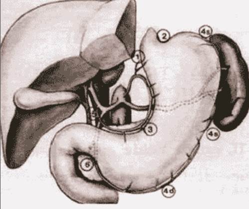 –ис. 1. Ћокализаци€ и нумераци€ лимфоузлов согласно J.R.S.G.C.
 –ис. 2. Ћокализаци€ и нумераци€ лимфоузлов согласно J.R.S.G.C.
1 - правые паракардиальные лимфоузлы;
2 Ц левые паракардиальные лимфоузлы;
3 Ц лимфоузлы малой кривизны;
4 Ц лимфоузлы большой кривизны;
4s Ц лимфоузлы вдоль левой желудочноЦсальниковой артерии и коротких желудочных сосудов;
4d Ц лимфоузлы вдоль правой желудочноЦсальниковой артерии;
5 Ц надпривратниковые лимфоузлы;
6 Ц подпривратниковые лимфоузлы;
7 Ц лимфоузлы левой желудочной артерии;
8 Ц лифоузлы общей печеночной артерии;
9 Ц лимфоузлы чревного ствола;
10 Ц лимфоузлы ворот селезенки;
11 Ц лимфоузлы селезеночной артерии;
12 Ц лимфоузлы печеночноЦдвенадцатиперстной св€зки;
13 Ц лимфоузлы позади поджелудочной железы;
14 Ц лимфоузлы корн€ брыжейки;
15 Ц лимфоузлы вдоль средней ободочной артерии;
16 Ц парааортальные лимфоузлы;
110 Ц нижние параэзофагеальные лимфоузлы;
111 Ц диафрагмальные лимфоузлы.
”словно вышеприведенные лимфоузлы формируют 4 этапа метастазировани€. ѕервый этап (N1): лимфоузлы св€зочного аппарата желудка (1Ц6). ¬торой этап (N2): лимфоузлы левой желудочной (7), общей печеночной (8), чревного ствола (9), в воротах селезенки (10), вдоль селезеночной артерий (11). “ретий этап (N3): лимфоузлы гепатодуоденальной св€зки (12), ретропанкреатодуоденальные лимфоузлы (13), корн€ брыжейки поперечноободочной кишки (14). „етвертый этап (N4): лимфоузлы вдоль средней ободочной артерии (15), парааортальные (16). ¬овлечение лимфоколлекторов N1ЦN2, а также 12 группы (гепатодуоденальной св€зки) рассматриваетс€ как регионарное метастазирование, N3ЦN4 Ц как отдаленные метастазы. Ётапность метастазировани€ €вл€етс€ условной, так как дл€ разной локализации опухоли в желудке не €вл€етс€ идентичной. роме того, существуют так называемые Ђпрыгающиеї, Ђскачущиеї (skipping) метастазы, обнаруживаемые в непораженных промежуточных участках путей лимфооттока.
—огласно последней классификации по стадированию –∆ индекс “ означает не размер опухоли, а степень прорастани€ стенки желудка: T1 Ц прорастание слизистой и подслизистого сло€, “2 Ц прорастание мышечного сло€ до субсерозы, “3 Ц пенетраци€ серозы желудка, “4 Ц прорастание прилежащих структур. ‘ормально, тотальный рак желудка с прорастанием всех слоев (но не прорастающий в соседние структуры) и небольша€ опухоль размерами до 2 см с выходом на серозу могут попасть в одну стадию, что говорит о том, что даже последн€€ классификаци€ не €вл€етс€ идеальной. »ндекс N при установке стадии заболевани€ присваиваетс€ следующим образом: поражение лимфоузлов от 1 до 6 расцениваетс€ как N1, от 7 до 15 как N2, поражение метастазами более 15 лимфоузлов расцениваетс€ как N3. оличество пораженных лимфоузлов призвано помочь в объективизации стадировани€ –∆, а также, в известной мере, указывает на объем и адекватность операции. √астрэктоми€ D1 (от слова dissection) предусматривает удаление перигастральных лимфоколлекторов, расположенных в св€зочном аппарате желудка (N1Ц6), гастрэктоми€ D2 означает удаление кроме N1Ц6, удаление лимфоузлов чревного ствола (N9) и его ветвей Ц левой желудочной (N7), общей печеночной артерии (N8), селезеночной (N11), лимфоузлов ворот селезенки (N3). Ћимфодиссекци€ D3 предполагает в дополнение к выше перечисленным лимфоузлы гепатодуоденальной св€зки (N12), ретропанкреатодуоденальные (N13), лимфоузлы корн€ брыжейки (N14), брыжейки поперечноободочной кишки (N15), парааортальных лимфоузлов, расположенных на уровне брюшной аорты (N16). Ќа IV ћеждународном онгрессе по раку желудка в ЌьюЦ…орке (—Ўј, 2001 год) лимфодиссекци€ D2 определена, как стандартный объем радикального хирургического вмешательства, так как улучшает отдаленные результаты и снижает частоту местных рецидивов. —читаетс€, что при лимфодиссекции D2 должно удал€тьс€ не менее 27 лимфоузлов, при D3 Ц не менее 40 лимфоузлов. Ќа сегодн€шний день можно постулировать, что больные, которым при оперативном лечении не произведена лимфодиссекци€ D2, должны считатьс€ пациентами с неустановленной стадией и не должны включатьс€ (во избежании феномена Ђмиграции стадииї) в статистические отчеты и международные протоколы исследовани€.
Ѕолее 10 лет назад предложена эндоскопическа€ резекци€ слизистой при раннем –∆ (EMR Ц endoscopic mucosal resection). ѕоказани€ дл€ EMR могут быть сформулированы следующим образом (при отсутствии отдаленных метастазов): высоко и умереннодифференцированна€ аденокарцинома; макроскопически IIa тип роста согласно €понской классификации (superficial elevated type) размерами не более 2 см в диаметре или IIc тип (superficial depressed type) менее 1 см, отсутствие €звенных изменений в опухоли; поражение слизистой. ѕри поражении подслизистого сло€ необходима операци€ с лимфодиссекцией D2. ќчевидно, что дополнительным условием дл€ EMR €вл€ютс€ квалифицированный медперсонал и соответствующа€ аппаратура [18,26,27].
¬ хирургии иногда используют термин Ђусловно радикальна€ операци€ї, при котором подразумеваетс€ полное удаление опухоли и видимых ее про€влений, однако предполагаетс€ раннее метастазирование или наличие неудаленных метастазов. ≈сли во всех удаленных лимфоузлах обнаруживаютс€ метастазы рака, то €сно, что веро€тность оставленных метастазов крайне высока. ƒанное утверждение подтверждаетс€ тем фактом, что при выходе опухоли на серозный покров частота обнаружени€ метастазов в забрюшинных лимфоузлах достигает 15Ц35%. ѕоэтому лечебный эффект лимфодиссекции вправе можно ожидать в тех случа€х, когда удал€етс€ следующий за этапом метастазировани€ путь лимфооттока. “ак, при поражении уровн€ N1 (группа лимфоузлов 1Ц6) нужно проводить лимфодиссекцию D2, при поражении уровн€ N2 (группа 1Ц11) лечебный эффект ожидаетс€ от лимфодиссекции D3. ѕо мнению р€да хирургов, наиболее четко лечебный эффект лимфодиссекции D2 про€вл€етс€ при II и IIIa стадии. насто€щему времени четкие показани€ к лимфодиссекции D3 еще не определены.
|
|
|
омбинированные операции при раке желудка
ѕри прорастании опухоли желудка в соседние органы широко примен€ютс€ резекции вовлеченных структур (печени, толстой кишки, почки, поджелудочной железы, надпочечника, селезенки). ѕри отсутствии отдаленных метастазов в печени, по брюшине, Ђпакетовї региональных узлов могут выполн€тьс€ такие обширные операции, как гастропанкреатодуоденальна€ резекци€ [21]. ¬опрос об об€зательной спленэктомии при лимфодиссекции D2 также €вл€етс€ дискуссионным. —торонники спленэктомии считают, что без нее невозможно удаление лимфоузлов ворот селезенки, противники указывают на редкость метастазировани€ –∆ в указанную зону, например, при дистальном –∆, а также на увеличение риска панкреатического свища и поддиафрагмального абсцесса. јналогичные аргументы привод€тс€ и в случае резекции тела и хвоста поджелудочной железы при отсутствии €вных признаков врастани€ в поджелудочную железу. ѕри раках проксимального отдела, опухол€х задней стенки тела больше 2 см в диаметре большинство авторов производ€т спленэктомию. –оль спленэктомии как таковой на прогноз при –∆ изучаетс€. ѕри распространенных формах иногда провод€тс€ операции с резекцией артерий чревного ствола или его полной перев€зкой (операци€ Appleby), эвисцерации левого верхнего квадранта брюшной полости, включающей гастрэктомию, спленэктомию, субтотальную панкреатэктомию, резекцию поперечноободочной кишки, левостороннюю адреналэктомию (left upper abdominal evisceration) и т.д.
’ирургическое лечение кардиоэзофагеального рака
¬ большинстве стран ≈вропы заболеваемость раком кардиоэзофагеальной зоны растет. Ќа согласительной конференции ћеждународной ассоциации по –∆ и ћеждународного общества по заболеванию пищевода в 2000 году экспертной комиссией рекомендована классификаци€, предложенна€ немецким хирургом J.R. Siewert. ¬ основе ее Ц ориентаци€ на анатомический центр опухоли, расположенный относительно ZЦлинии, зоны перехода эпители€ пищевода в желудочный. ѕри этом используютс€ два хирургических доступа: чрезбрюшинный и левосторонний торакоабдоминальный. „астота обнаружени€ медиастинальных метастазов при раках кардии достигает 30%, а п€тилетн€€ выживаемость в таких случа€х не превышает 10% [21]. “ак как многие хирурги не отмечают лечебный эффект медиастинальной лимфодиссекции при метастатическом поражении средостени€, а также в св€зи с улучшением хирургического пособи€ с возможностью формировани€ Ђвысокихї трансдиафрагмальных эзофагоеюноанастомозов при лапаротомии, тораколапаротомный доступ больше не €вл€етс€ преимущественным при раке кардии. ќсобенно актуальным становитс€ вопрос о хирургическом доступе при раке кардиального отдела у пожилых, ослабленных больных с сопутствующими кардиореспираторными заболевани€ми.
ѕаллиативные операции при раке желудка
Ѕольные с €влени€ми стеноза выходного отдела, дисфагией, кровотечением из распадающейс€ опухоли, с пенетрацией опухоли в соседние органы и структуры, €влени€ми кишечной непроходимости (чаще при прорастании в поперечноободочную кишку), анемией, кахексией с обезвоживанием (особенно при дисфагии) часто попадают в общехирургические отделени€ и больницы терапевтического профил€. сожалению, даже сами по себе вышеперечисленные осложнени€, особенно у пожилых и пациентов старческого возраста в сознании многих врачей и хирургов ассоциируютс€ с крайней запущенностью процесса и неоперабельностью. ”величение доли пожилых людей в –оссии только повысит актуальность проблемы. ¬месте с тем практически все перечисленные осложнени€ встречаютс€ либо при небольших опухол€х, когда возможна радикальна€ операци€, либо при местнораспространенных опухол€х, при которых также возможно оперативное пособие. Ќемаловажную роль играет и так называемый Ђчеловеческий факторї. “ак, по данным –ќЌ÷ –јћЌ им. Ќ.Ќ. Ѕлохина, у половины больных, которым выполнена Ђпробна€ї лапаротоми€ в неспециализированных, чаще в общехирургических клиниках, в последующем в специализированном онкологическом учреждении выполн€лось хирургическое пособие. —реди паллиативных операций, нар€ду с общеизвестными (гастрэктоми€, еюностома, обходные гастроэнтероанастомозы при раках, локализующихс€ в антральном отделе), следует отметить Ђшунтирующиеї операции, которые выполн€ютс€ чаще при неоперабельном раке проксимального отдела и кардиоэзофагеальной зоны. —уть операции заключаетс€ в обходном эзофагоеюноанастомозе, который может быть выполнен как чрезплевральным, так и абдоминальным доступом, что освобождает больного от мучительной дисфагии и необходимости пользоватьс€ еюностомой.
’имиотерапи€ при раке желудка
–ак желудка мало чувствителен к химиотерапии. ƒо сих пор не освещен вопрос, почему у одних больных метастазами поражаетс€ только печень при интактных забрюшинных лимфоузлах и брюшины, у других же при Ђчистойї печени Ц бурное метастазирование по брюшине и в лимфоузлы. ќчевидно, что речь идет о разной биологической активности опухолевых клеток. ¬озможно, последние различаютс€ в различной экспрессии молекул, ответственных за тропность либо к мезотелию брюшины, либо к эндотелию сосудов печени, лимфоузлов и т.д. “ем не менее выход опухоли на серозную оболочку желудка считаетс€ независимым фактором метастазировани€ по брюшине. Ёффективность химиотерапии при –∆ не превышает 30Ц40%. ¬ большинстве стран примен€ютс€ комбинации PF (цисплатин и 5Цфторурацил), ELF (этопозид, кальци€ фолинат и 5Цфторурацил), ECF (эпирубицин, цисплатин и 5Цфторурацил). ѕри этом пон€тие Ђэффективностьї часто включает достаточно разнородные пон€ти€: субъективный эффект, объективный эффект Ц уменьшение опухоли или метастазов, общую или безрецидивную выживаемость и т.д. ¬ целом считаетс€, что применение химиотерапии улучшает качество жизни, то есть оказывает субъективный эффект, увеличивает безрецидивную выживаемость, не вли€€ на общую выживаемость, особенно при радикальных операци€х, мало эффективна в адъювантном режиме и в р€де случаев увеличивает продолжительность жизни при неоперабельном –∆. –€д исследований в японии и орее показал эффективность адъювантной внутрибрюшной химиотерапии при прорастании опухоли серозного покрова. ак модификаци€ указанного метода примен€етс€ внутрибрюшна€ гипертермическа€ химиотерапи€. Ёффективность метода показана как при наличии метастазов на брюшине, так и с точки зрени€ профилактики последних. Ђ«олотым стандартомї в лечении –∆ последние 10 лет считалась комбинаци€ с включением цисплатина, фторурацила и кальци€ фолината.
Ќа последнем конгрессе ASCO в 2003 году в качестве препаратов, показавших эффективность в двух рандомизированных исследовани€х, названы иринотекан и доцетаксел.
«аключение
–ак желудка в –оссии остаетс€ чрезвычайно острой проблемой. —мертность за последние годы в нашей стране не снизилась. ¬ы€вл€емость ранних форм, с одной стороны, низка€, однако, с другой стороны, €вл€етс€ единственным шансом на выздоровление. линические про€влени€ раннего –∆ не €вл€ютс€ патогномоничными, но часто скрываютс€ под обычными Ђжелудочнымиї жалобами. —крининговые программы, проводимые в экономически развитых странах, €вл€ютс€ дорогими. ¬ –оссии требуетс€ национальна€ скринингова€ программа по вы€влению наиболее распространенных онкологических заболеваний, а в услови€х современной действительности скрининг –∆ должен проводитьс€ хот€ бы в группах фоновых и предраковых заболеваний. ¬ этой св€зи требуетс€ попул€ризаци€ знаний (включа€ телевидение, радио, распространение буклетов и т.д.) о –∆ врачей общего профил€ и среди населени€. ќсоба€ роль должна отводитьс€ терапевтам, гастроэнтерологам, эндоскопистам. ƒейственной мерой профилактики –∆ может служить изменение образа питани€. Ѕольные –∆ должны лечитьс€ в специализированных учреждени€х. ѕри семейном –∆ должно проводитьс€ медикогенетическое консультирование родственников. Ђ«олотым стандартомї в хирургическом лечении €вл€етс€ гастрэктоми€ (резекци€) с объемом лимфодиссекции D2.
Ћ»“≈–ј“”–ј
1. ћ.Ќ.ƒавыдов, –ак проксимального отдела желудка: стандарт хирургического лечени€, основанный на 30-летнем опыте / ћ.Ќ.ƒавыдов, ћ.ƒ. “ер-јванесов, ».—.—тилюди, ј.Ѕ.√ерманов, ќ.Ќ.≈фимов и др. / ¬естник –оссийской јкадемии ћед. наук, ћ.: ћедицина, - 2002, - є1, - —. 25-28.
2. Crew K.D., Neugut A.I. Epidemiology of gastric cancer // World J Gastroenterol. Ц 2006. Ц Vol. 12, є3. Ц P. 354-362.
3. Maconi G., Manes G., Porro G.B. Role of symptoms in diagnosis and outcome of gastric cancer // World J Gastroenterol. Ц 2008. Ц Vol. 14. Ц є8. Ц P. 1149-1155.
4. Rothenberg M.L. »ринотекан (—–“Ц11): новые данные и перспективы применени€ Ц колоректальный рак и другие злокачественные опухоли. The Oncologist, 2001:6;66Ц80.
5. Parkin D.M. Global cancer statistics in the year 2000 // Lancet oncol. - 2001.- vol. 2 Sept. - P. 533- 543.
6. Hamashima C., Shibuya D., Yamazaki H. et al. The Japanese guidelines for gastric cancer screening // Jpn J Clin Oncol. Ц 2008. Ц Vol. 38. Ц є4. Ц P. 259-267.
7. Kaise M., Kato M., Urashima M. et al. Magnifying endoscopy combined with narrow-band imaging for differential diagnosis of superficial depressed gastric lesions // Endoscopy. Ц 2009. Ц Vol. 41, є4. Ц P. 310-315.

