


Anaphase I. Homologous chromosomes migrate away from each other, going to opposite poles. Each chromosome still consists of two chromatids.
Telophase I. Telophase I is similar to telophase of mitosis. The chromosomes reach the opposing poles and cytokinesis occurs, giving rise to two daughter cells. Each cell possesses 23 chromosomes, the haploid (1n) number, but because each chromosome is composed of two chromatids, the DNA content is still diploid. Each of the two newly formed daughter cell enters meiosis II.
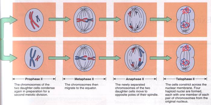
Meiosis II (Equatorial Division)
The equatorial division is not preceded by an S phase. It is very much like mitosis and is subdivided into prophase II, metaphase II, anaphase II, telophase II, and cytokinesis. The chromosomes line up on the equator, followed by the chromatids migrating to opposite poles, and cytokinesis divides each of the two cells, giving a total of four daughter cells from the original diploid germ cell. Each of the four cells contains a haploid amount of DNA content and a haploid chromosome number.
Unlike the daughter cells resulting from mitosis, each of which contains the diploid number of chromosomes and is an identical copy of the other, the four cells resulting from meiosis contain the haploid number of chromosomes and are genetically distinct because of reshuffling of the chromosomes and crossing over.

Cell death.
There are two forms of cell death: necrosis and apoptosis. Necrosis is caused by various external factors, chemical or physical, which influence the cell directly or indirectly and cause its death. Apoptosis is the programmed cell death.
Theme: The Nucleus of the cell.
1. The functions of the Nucleus.
2. Nucleus structure of interpfase cell.
3. The structure and functions of Nuclear Pores.
4. The structure of Interpfase Chromosome.
5. The structure of Nucleolus.
Theme: Reproduction of the cells.
Aging and death of cells.
1. Cell cycle definition.
2. Stages of interphase, characteristic features:
a) postmitotic period G1;
b) synthetic period S;
c) premitotic period G2.
3. Stages of mitosis:
a) prophase;
b) metaphase;
c) anaphase;
d) telophase.
4. Characteristic of polyploidy and endoreduplication.
5. Meiosis peculiarities:
- reductional division;
- equatorial division.
6. Aging and death of cells. Necrosis and apoptosis.
Practical pat.


 1 3
1 3
 |
Slide №9 Cell with a round form of the nucleus.
Staining: hematoxylin – eosin.
1 – nucleus; 2 – cytoplasm; 3 - nucleolus.

 1 2
1 2

Slide №10 Сell with a rod like nucleus.
Staining: hematoxylin – eosin.
1 – nucleus; 2 - cytoplasm.

 |
 |
Slide №11 Cell with a segmented nucleus.
Staining: hematoxylin – eosin.
1 – cytoplasm; 2 – nucleus may have three to seven lobes connected by thin strands of nucleoplasm.

Slide №12 Mitosis.
Staining: iron hematoxylin.
1 – interkines; 2 – early prophase; 3 – late prophase; 4 – metaphase; 5 – microtubules of spindle; 6 – anaphase; 7 – telophase.

Diagram №5 Structure of nuclear pores.
1 – perinuclear space; 2 – an inner membrane of a nuclear envelope; 3 - an outer membrane of a nuclear envelope; 4 – peripheral granules; 5 – central granule; 6 - fibrillar –globular molecules; 7 – diaphragm of pore; 8 – fibrils of chromatin.

Diagram №6 Mitosis.
1 – prophase; 2 – metaphase; 3 – anaphase; 4 – telophase.


Diagram №7 Meiosis.
Electron Micrographs.
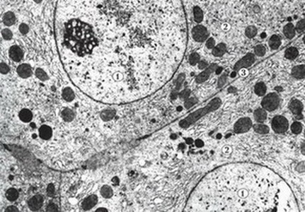
Fig. 1.
Tightly attached cells of the liver (hepetocytes).
1. Nucleus.
2. Cytoplasm
3. Cell membranes of adjacent cells and narrow interecellular space.

Fig. 2.
Round – shaped cell (lymphocyte).
1. Nucleus.
2. Cytoplasm.
3. Plasmolemma.

Fig. 3.
Goblet cell (exocrinocyte).
1. Nucleus.
2. Cytoplasm with the secretory granules.
3. Plasmolemma.
4. Basal pole.
5. Apical pole.

Fig. 4.
Cell with the processes (neuron).
1. Nucleus.
2. Cytoplasm.
3. Plasmolemma.
4. Process.

Fig. 5.
Columnar cells (absorptive cells).
1. Nucleus.
2. Cytoplasm.
3. Plesmolemma.
4. Microvilli on the apical pole.

Fig.6.
Thrombocytes.
1. Plasmolemma.
2. Cytoplasm.

Fig. 7.
Multinuclear structure – symplast (chorionic villi).
1. Nucleoli.
2. Plasmolemma.
3. Cytoplasm.

A
Fig. 8.
Electron image of plasmolemma (trilaminar structure).
A – free surface of the cel:
1. Inner dense layer.
2. Inter – mediate electron lucid layer.
3. Outer dense layer.
4. Glucocalyx.
5. Cytoplasm.

Fig. 9.
Phagocytosis.
1. Cell’s cytoplasm.
2. Projection of the cytoplasm.
3. Invagination of the cell membrane.
4. Phagocytic material.

Fig. 10.
Macropinocytosis ba microvilli of endothelial cells of blood capillary.
1. Cytoplasm of endothelial cell.
2. Lumen of capillary.
3. Microvillus.
4. Vacuole.

Fig. 11.
Micropinocytosis in endothelial cells of blood capillary.
1. Plasmolemma of endothelial cell.
2. Caveolae.
3. Pinocytotic vesicles.
4. Cytoplasm of endothelial cell.
5. Lumen of capillary.

Fig. 12.
System of intercellular junctions.
1. Digital junction.
2. Digital cellular lock.
3. Tight junction (zonula occludens).
4. Desmosome.

Fig. 13.
Intercalated disk. Connection between cardiac muscle cells.
1. Plasmolemma.
2. System of desmosomes and gap junctions (nexuses).
3. Myofibrils.
4. Mitochondrion.

Fig. 14.
Specialized connection between nervous cells - synapse.
1. Presyneptic pole.
2. Postsynaptic pole.
3. Synaptic cleft.
4. Syneptic vesicles.

Fig. 15.
Rough endoplasmic reticulum.
1. Cisternae.
2. Ribosomes.
3. Fragment of the nucleus.

Fig. 16.
Smooth endoplasmic reticulum.
1. Fragment of the nucleus.
2. Cisternae of the endoplasmatic reticulum.
3. Mitochondrion.
4. Liposome.

Fig. 17.
Golgi complex.
1. Cysternae.
2. Vesicles.
3. Vacuoles.
4. Fragment of the nucleus.

Fig. 18.
Mitochondrion.
1. Outer membrane.
2. Inner membrane.
3. Crictae.
4. Mitochondrial matrix.
5. Hyaloplasm.

Fig. 19.
Lysosomes.
1. Primary lysosome.
2. Secondary lysosome (autophagosome).
3. Residual body.

Fig. 20.
Myofibrils in the cytoplasm of the cardiac muscle cell.
1. Miofilaments of myofibrils.
2. Mitochondria.

Fig. 21.
Cilia (ciliated columnar cells apical surface of the mucus epithelial lamina of nasal cavity).
1. Apical pole of the cell.
2. Cilia.
3. Basal corpuscle.

Fig. 22.
Inclusions of glycogen in hepatocytes (liver).
1. Glycogen granules.
2. Fragment of the nucleus.
3. Mitochondrion.
4. Rough endoplasmic reticulum.

Fig. 23.
Inclusion of lipid in cytoplasm of the hepatocytes.
1. Lipid droplets.
2. Fragment of the nucleus.
3. Mitochondria.

Fig. 24.
Secretory inclusions in pancreatic acinar cells (pancreas).
1. Nucleus.
2. Cytoplasm.
3. Secretory inclusions in the apical pole of the cell.

Fig. 25.
Round – shaped nucleus.
1. Nucleoplasm.
2. Cytoplasm.
3. Nuclear envelope.
4. Nucleolus.

Fig. 26.
Fragment of the nucleus.
1. Nucleoplasm.
2. Cytoplasm.
3. Outer membrane of the nuclear envelope.
4. Inner membrane of the nuclear envelope.
5. Nuclear pore.

Fig. 27.
Structure of the nucleolus.
1. Pars fibrosa.
2. Pars granulosa.
3. Nucleolar associated heterochromatin.
4. Nucleoplasm.
5. Nuclear envelope.

Fig.28.
Intercellular substance
(loose connective tissue).
1. Cell (fibroblast).
2. Intercellular substance:
a) fibrous elements;
b) amorphic component.
Questions to modul.
1. The structure of the Plasmolemma.
2. Characteristic of receptive and transport functions of the plasmolemma.
3. The structure of intercellular contacts: Simple contact, Zonular occludentes, Synapse.
4. The structure of intercellular contacts: Desmosome, Zonular adherents, Gap junctions.
5. Characteristic and structure of symplast and sincytium.
6. The structure and functions of Mitochondria.
7. The structure and functions of Lysosomes and Peroxisomes.
8. The structure and functions of Agranular Endoplasmic Reticulum.
9. The structure and functions of Granular Endoplasmic Reticulum and Ribosomes.
10. The structure and functions of Golgi Bodies.
11. The structure and functions of Microfilaments and Microtubules.
12. The structure and functions of Cytocentrum (centrosome).
13. The structure and functions of Cilia and Flagella.
14. Name of types Inclusions and their functions.
15. Reproduction of cells definition.
16. Cell cycle definition.
17. Stages of interphase, characteristic features.
18. Stages of mitosis.
19. Characteristic of polyploidy (endoreduplication).
20. Meiosis peculiarities.
21. Aging and death of cells. Necrosis and apoptosis.
LECTURE № 1
Topic: Introduction into embriology. Sex cells. Fertilization.
Embryogenesis is the process of development of an organism from fertilization till birth or way out from ovular membranes.
Embryologia is the science studying both general regularities of embryogenesis and peculiarities processes of individual development of an organism.
Tasks of medical embryology.
They are:
1) to study mechanisms of embryo development;
2) to understand the defects in embryo development;
3) purposeful influence on embryo development.
Main stages of human embryogenesis.
Early: Late:
1. Fertilization. 1. Histogenesis.
2. Cleavage. 2. Organogenesis.
3. Gastrulation (early, late).
4. Formation of axial complex of organs.
5. Formation of embryo body.
Fertilization
The development of multicellular organism begins from one cell, called zigote. The zygote appears as a result of fusion of two parent sex cells into one. This process is called fertilization. Male and female sex cells differ both morphological features and physiological properties.

 Female sex cell (ovum, oocyte)
Main features.
1. Ovum is much larger than somatic cells. It is a cell of about 130 – 140 micrometres and somatic cells are only 10 – 20 mcm.
2. It has much cytoplasm (to provide the embryo with the initial cytoplasm mass).
3. In cytoplasm all the necessary organelles but centrosome (cell centre) no are present. So it can’t divide.
4. It has haploid set of chromosomes: 22 autosomes and 1 sex – X chromosome.
5. Presence of nutrients as yolk granules. Yolk is the compound of protein and lipids. The egg cells are divided into following types according to yolk amount:
Alecithal. Isolecithal.
(Without yolk) (Little yolk)
They don’t contain Yolk granules are distributed
nutrient reserve. They evenly through the cytoplasm:
are typical for inverterbrates a) primary isolecithal typical for
which conduct a parasitic way animals, which aquire early the
of life (echinoccus, tapeworm, ability for original feeding-lancet helminthes) b) secondary isolecithal are typical for animals with antenatal
development – mammals, the man.
Telolecithal
(contain a large amount of yolk)
a) Moderately telolecithal. Yolkis distributed on one pole-amphibian (frog).
b) Centrolecithal. Yolk is in the centre and nucleus on the periphery - insects.
c) Sharply telolecithal. There is much yolk, it is distributed through almost the whole cell – birds, reptile.
6. Presence of cortical granules,which are spread along the periphery of cytoplasm and consist of proteoglycans and glycoproteins.
7. An egg cell is covered with ovolemma and has external layers.
a) primary, which is the product of cortical granules – zone pellucida.
It is composed of glycoproteins and its functions are:
- protection from mechanical injuries;
- barrier, which allows the spermatozoon of only its own species pass through this layer.
b) secondary – granular zone or corona radiata, consists of follicular cells. Its functions are:
- defensive;
- trophic
8. Egg cells are immobile.

1) nucleus; 2) granular endoplasmic reticulum; 3) cytoplasm; 4) mitochondria; 5) Golgi bodies; 6) yolk granules; 7) cortical granules; 8) microvilluses; 9) zone pellucida; 10) corona radiata.
Male sex cell (spermatozoon).
Main features.
1. The concentration of spermatozoon number is 300 million in ejaculate (one portion). A single ejaculate normally contains approximately 50 to 100 million spermatozoa per milliliter. A man whose sperm count is less than 20 million spermatozoa per milliliter is considered sterile.
2. It has a specific form – filament.
3. They are mobile.
4. They are very small; it long about 60 micrometer.
Asingle ejaculate normally contains approximately 50 – 100 million spermatozoa per milliliter. A man whose sperm count is less than 20 million spermatozoa per milliliter is considered sterile.
A spermatozoon consists of the head, neck and tail.
In the head there is a nucleus with haploid set of chromosomes: 22 autosomes and 1 sex chromosome. It may be X or Y. So, spermatozoons are divided into two types: 1) (22a + Y) give rise to male organism; 2) (22a + X) give rise to female organism.
The acrosome is located over the nucleus in the head. It has originated from Golgi apparatus. Acrosome contains hydrolythic enzymes: tripsin and hyaluronidase, which are capable to dissolve the ovum membranes at the moment of fertilization.
A thin cytoplasm layer, covering the nucleus and acrosome of the head, continues to the neck. The neck contains a proximal centriole; a distal centriole is beneath it. The axoneme beginning from distal centriole is the principal part of the tail. It is the movement apparatus.
The tail segment of a spermatozoon consists of the first part, the main part and the end part. In the first part there are mitochondria, which are located coil-like around the axoneme. They provide the spermatozoon with energy to move. The main part of the tail contains the axoneme and is covered a narrow layer of cytoplasm which disappears in the end part. The аxoneme is composed of two central microtubules and 9 doublet of peripheral ones. The movement speed of spermatozoon is 50 micrometer per second.
 I. Head. II. Neck. III. Tail.
1) acrosome; 2) nucleus; 3) proхimal centriole; 4) distal centriole; 5) mitochondria;
6) axoneme.
Spermatozoon preserve the ability to fertilization in the female sex organs for two days. Off all organelles spematozoons contain only acrosome, mitochondria and 2 centrioles (centrosome).
Spermatozoon functions:
1. It transmit genetic information during fertilization.
2. It start up the programme of further development in the zygota.
5. It determines the sex of new individual (X - carrying sperm will produce a female embryo, Y – carrying sperm will produce a male embryo).
Fertilization is the fusion process of two sex cells. It occurs in the uterine tube and consists of two phases – distant phase and contact phase.
During the distant phase the process of capacitation occurs. The enzymes, which are produced in uterine glands, erode glycoprotein covering of spermatozoon. They become mobile. This occurs in the female sex tracts. Sperm release androgamones, which are biologically active substances.
They deprive the female sex cell the ability to move. At the same time follicular cells which surround the female egg cell release gynogamones. These substances activate spermatozoons and attract them to themselves. This ability of sperms to move in the direction of increasing the concentration gynogamones is called chemotaxis. The sperm ability to move against fluid current is called reotaxis.
A lot of spermatozoons reach the female sex cell (ovum), surround it, make it immobile and sperm which has maximum ability, goes between follicular cells and the first adheres to zone pellucida. At this time phase of contact interaction begins. Spermatozoon adhere to zone pellucida receptors; after that acrosomal reaction begins. As a result of acrosomal reaction release of hyaluronidase and tripsin enzimes from spermatozoon acrosome takes place. These enzymes erode zone pellucida in the point of contact of spermatozoon with it. The content of the head and neck (nucleus and centriole) penetrate in the female sex cell the hole in the point of contact of acrosome and zone pellucida.
After penetration of spermatozoon cortical reaction starts. The out low of cortical granules content occurs. This results in zone pellucida swelling. It formd fertilization membrane and follicular cells are seperated from fertilization membrane. As a result another sperm cannot penetrate the ovum and thus monosperm fertilization occurs. Both nucleus, of male and female sex cells, located in the ovum cytoplasm, swell, chromatin in them loosen, and they get the name pronucle, and a short period of their existence – syncaryon. Male pronuclus comes nearer to the female one and fuses. This result in formation of one cell - zygote. Zygote has diploid set of chromosomes, which includes genes, inherited from both parents. During this period reconstruction in cytoplasm and nuclues takes place. It determines following processes of development.

Diagram Fertilization: 1) cytoplasm; 2) nucleus; 3) zone pellucida; 4) corona radiate; 5) head of spermatozoon; 6) neck of spermatozoon; 7) tail of spermatozoon; 8) penetrate of spermatozoon into the female sex cell; 9) membrane of fertilization; 10) pronucleus of the female sex cell; 11) pronucleus of the male sex cell; 12) centrosome; 13) syncarion.
Theme: Embryology. Sex cells.
Fertilization.
1. Embryogenesis, embryologia definition.
2. Main stages of human embryogenesis.
3. Main features of the female sex cell.
4. Oocytes types.
5. Structure of the ovum.
6. Main features of the male sex cell.
7. Structure of the spermatozoon.
8. Fertilazation, stages and biological significance.


Slide №8

Slide № 7 Oocyte.
Staining: hematoxylin – eosin.
1 – follicular epithelium; 2 – corona radiata; 3 – zona pellucida; 4 – cytoplasm;
5 – nucleus with nucleolus.

Diagram №9 Structure of ovum.
1 - nucleus; 2 - granular endoplasmic reticulum; 3 - cytoplasm; 4 - mitochondria; 5 - Golgi bodies; 6 - yolk granules; 7 - cortical granules; 8 - microvilluses; 9 - zone pellucida; 10 - corona radiata.

Diagram №2 Sructure of spermatozoon.
I. Head. II. Neck. III. Tail.
1- acrosome; 2 - nucleus; 3 - proхimal centriole; 4 - distal centriole; 5 - mitochondria;
6 - axoneme.

Diagram №3 Fertilization.
1 - cytoplasm; 2 - nucleus; 3 - zone pellucida; 4 - corona radiate; 5 - head of spermatozoon; 6 - neck of spermatozoon; 7 - tail of spermatozoon; 8 - penetrate of spermatozoon into the female sex cell; 9 - membrane of fertilization; 10 - pronucleus of the female sex cell; 11 - pronucleus of the male sex cell; 12 - centrosome; 13 - syncarion.
LECTURE № 2
Topic: Cleavage. Gastrulation.
Cleavage.
Cleavage is the division of the zygote, in the result of which multicellula organism - blastula - is formed. Cleavage occurs by means of mitosis. This is a very short period of interphase between two mitosis and it begins with S phase of the cell cycle. G phase of the cell cycle, during which new formed cells are growing, is absent. Such division results in size decrease of dividing cells half, and as a result, the blastula size does not exceed the zygota size. Cells, from which the embryo is constituted during the cleavage are called blastomeres.
Different types of animals have their own mode of cleavage. It depends on yolk amount in the egg cell, as yolk prevents the cleavage.
Modes of cleavage
 | |
 Complete Incomplete

Holoblastic (the egg cell is Meroblastic (the egg cell is divided completely) divided partly)
|  |
Regular Irregular
Blastomeres are the The size of blastomeres are
same size different: microblastomeres,
macroblastomeres
Synchronous Asynchronous
Blastomeres are divided Blastomeres are divided
simultaneously not simultaneously
Cleavage is completed by blastula formation. Blastula is a multicellular monolayer embryo. It consists of blastula wall - blastoderm, which is composed of cells - blastomeres. There is a cavity - blastocell inside blastule:


 1
1  2
2
1) blastoderm; 2) blastomeres; 3) blastocell.
The cleavage peculiarities in the human. Chronology of the process.
In thirty hours after fertilization the zygote, which is in the uterine tube enters the cleavage. For the human zygote complete and asynchronous cleavage, that is such cleavage during, which some blastomeres are divided more often, others - more seldom. That is blastomeres divided with different speed. After the stage of two blastomeres the stage of three blastomeres comes and so on. A group of cells, consisting of 12 - 16 blastomeres is called morula. At the stage of 16 blastomeres all cells of morula are totipotent. This means that any blastomere can become the starting-point structure for development of the organism, as a whole 16 separate twins may develop. On the fourth day after fertilization the number of blastomeres increases up to 50 - 60 and blastula is formed. For the human the type of blastula - blastocyste – is typical. Blastocyste takes the form of vesicle. The transparent tunic is dissolved. If dissolving of the transparent tunic does not occur blastocyste won’t be able to the surface of attach uterus endometrium.
During the first four days the germ is in the cavity of uterine tube and move, with the fluid current and under the influence of peristaltic contraction into the uterine cavity. This is the tubal period in the development of the germ. The germ as free blastocyste is in the uterine cavity from the fifth till seventh day.

Diagram Fertilization and the fist 6 days of development.
1) ovary; 2) ovulation; 3) fertilization; 4) zygote; 5) uterine tube; 6) two cell stage;
7) four cell stage; 8) morula; 9) blastocyst; 10) cavity of uterus.
During the period (from the first till seventh day) nutrition is carried out partly by oocyte nutrients, partly by secretion of uterine tube and endometrium glands and is called vitelotrophic.
On the seventh day implantation comes. At this time blastocyst consists of 107 blastomeres, from which 69 form trophoblast, 8 - embryoblast, and 30 groups around embryoblast and form the inner cell mass. Under the influence of secretion of glycoproteins the membrane of fertilization is eroded. Blastocyst gets into contact with uterus endometrium.


2 3
1) embryoblast; 2) blastocell; 3) trophoblast.
1. The phase of adhesion - adhesion or attachment of blastocyst between excretory ducts of uterine glands.
2. Invasion phase - penetration of blastocyst into uterine mucosa with the help of proteolytic enzymes, which are produced by trophoblast. First epitheliocytes are eroded, then the connective tissue and, at last, the walls of vessels of endometrium. Hollow is formed in which blastocyst is located and then the hollow overgrown by the connective tissue. Prolifiration of endometrium epitheliocytes eliminates the defect of epithelium.


Diagram Implantation.
A – Adhesion B - Invasion
1) wall of the uterus; 1) endometrium with blood vessels and glands;
2) inner cell mass; 2) syncytiotrophoblast;
3) trophoblast; 3) cytotrophoblast;
4) blastocyst cavity; 4) inner cell mass;
5) cavity of the uterus. 5) embryonic entoderm;
6) lumen of uterus.
From the seventh day and until getting of trophoblast in contact with mother’s blood (the end of the fourth week), histiotrophic period of embryogenesis continues. Embryo’s nutrition is provided by secretion of uterine glands and erosion products of endometrium tissues by trophoblast.
From the moment of embryo’s contact with mother’s blood and untill newborn’s birth (from the second till ninth month) haematotrophic period continues. During this period provision of the embryo and fetus with nutritious substances and gas exchange take place by mother’s blood.
Trophoblast cells are actively divided and it turns from monolayer into bilayers. The internal layer has cellular structure and is called cytotrophoblast. The external layer does not have cellular structure and is called syncytiotrophoblast.
Gastrulation
Gastrulation is the stage of embryogenesis when three germ layers are formed: ectoderm,endoderm and mesoderm.
During gastrulation:
1) division and growth cells;
2) translocation of cells;
3) differentiation of cells take place.
Gastrulation is divided into two phase: early and late. During early gastrulation formation of ectoderm (external) and entoderm (internal) germ layers take place. During late gastrulation formation of mesoderm (middle germ layer) and axial germs of organs complex: neural tube, chordomesodermal germ and primary intestine.
Ways of gastrulation.
1. Invagination is observed at simple chordata. The bottom of blastula into blastocele is invaginated and two germ layers are formed: ecto- and entoderm.

2. Epiboly is characteristic for frogs. Small blastomeres are quickly divided and surround large blastomeres.

3. Delamination is characteristic for birds, mammalia and the man. Division or split of blastomeres into two layers external (ectoderm) and internal (entoderm) take place.

4. Immigration is characteristic for birds and the man. Migration of part of blastomere into blastocele ocurs.

Early gastrulation.
The period of early gastrulation is from the seventh to fourteenth day. By delamination division or split of embryoblast into epiblast and hypoblast takes place. At the same time cavitation or dissolution of internal cellular mass, which surrounds embryoblast, occurs. The cells of epiblast and hypoblast are divided.
Epiblast cells go upwards connect and form amniotic vesicle. The edges of hypoblast go downwards and form yolk sac. The bottom of amniotic vesicle is formed by epiblast and the rest parts of it form extraembryonic ectoderm. The roof of yolk sac is formed by hypoblast and from the rest part of it extraembryonic entoderm is formed. The bottom of amniotic vesicle and the roof of yolk sac formed embryonic disk.

Diagram Embryo at day 9
1) wall of uterus; 2) layers from trophoblast; 3) amnion; 4) amniotic sac cavity;
5) epiblast; 6) hypoblast; 7) bilaminar embryonic disc; 8) yolk sac cavity.
From hypoblast only extraembryonic entoderm is formed, and from epiblast embryo and extraembryonic ectoderm develop. Cells of germ disk begin to divide and move into the cavity of blastocyte and form extraembryonic mesoderm. These cells move in three currents. The first current surround the amniotic vesicle and form the external wall of amnion; the second current forms the external wall of yolk sac, and the third - attaches to trophoblast and participates in formation of chorion internal wall.
So, in man extraembryonic organs - chorion, amnion and yolk sac become the most developed during early gastrulation. Amnion wall is composed of extraembryonic mesoderm and extraembryonic ectoderm. Yolk sac wall is composed of extraembryonic mesoderm and extraembryonic entoderm. Chorion wall is composed of 3 layers: external - syncytiotrophoblast, middle - cytotrophoblast, internal - extraembryonic mesoderm.
Amniotic vesicle is attatched to chorion with the help of amniotic stalk, formed from extraembryonic mesoderm. Further, on the place of amnionic stalk, umbilical cord will be developed.
1. Syncytiotrophoblast
2. Cytotrophoblast
3. Extraembryonic mesoderm
4. Chorion
5. Amnion stalk
6. Extraembryonic mesoderm
7. Extraembryonic ectoderm
8. Amnion
9. Epiblast
10. Hypoblast
11. Germ disk
12. Extraembryonic entoderm
13. Extraembryonic mesoderm
14. Yolk sac


Diagram 6. Transverse section through the embryonic disc at the primitive streak
1 – primitive streak;
2 – hypoblast;
3 – yolk sac lining;
4 – mesoderm;
5 – cut edge of amnion;
6 – primitive streak;
7 – primitive node;
8 – epiblast (will become ectoderm).
Late gastrulation begins on the 14-15 day. Epiblast cells are intensively divided and imigrate from the head end to the caudal one in two currents. Here they meet and turning in the opposite direction, form thickened cellular band along middle line of germ disk - primary streak. On the anterior part of primary stripe cellular thicking looking like tubercle - primary knot - is formed.
From the cells of the anterior part of primary stripe embryonic entoderm is formed, from the cells of middle part of primary steak embryonic mesoderm is formed, and from the cells of the posterior part form extraembryonic mesoderm. Cells of primary knot and site of epiblast, lying before it, move into formed pit on the place of primary streak, extending in the form of cellular band - chorda.
1. Head end.
2. Caudal end.
3. Primary stripe.
4. Primary knot.
5. Formation of embryonic entoderm.
6. Formation of embryonic mesoderm.
7. Formation of extraembryonic mesoderm.
8. Mesoderm wings.
9. Neural tube.
10. Neural crest.
11. Dermal ectoderm.
Embryonic ectoderm is formed owing to those epiblast cells, which stay in their own place. Space between embryonic layers is filled with embryonic connective tissue - mesenchima, which is mainly formed from mesoderm. From 17-20 days the formation of axial germ of organs is completed. Just after the formation, the cells of chorda they are induce the ectoderm cells overlying it.
This cause their prolifiration, formation of neural plate, its caving in towards chorda. This results in the formation of neural groove and neural elevations. These elevations fuse and formation of neural tube begin in the neck region of embryo and continues in the caudal direction. Further, on the head end of neural tube enlargements - cerebral vesicles are formed.
Cells, from which neural crest is formed, are located between neural tube and ectoderm. Further, from neural crest spinal ganglia are formed. The process of neurulation is - process of neural tube formation.

Diagram 8. Neurulation
1 – level of section;
2 – surface ectoderm;
3 – neural plate;
4 – neural folds;
5 –neural drove;
6 – neural crest;
7 – surface ectoderm;
8 – neural tube.
Simultaneously (at the same time) differentiation of mesoderm occurs. On the 20 day of embryonic development somitic period begins. Dorsal sites of mesoderm, which lie on both sides of chorda, begin to divide into separate segments - somites. The process of segmentation and the formation of somites are taking place from the head towards the cauda of embryo. By the 35 day 44 pairs of somites are formed. Every somite differentiates into three parts, from the external part – dermatome - the source of connective tissue of skin (derma) is formed, from the central part – myotome - the source of skeletal muscular tissue, from internal part – sclerotome - the source of cartilaginous and osseous tissues.
Ventral mesoderm is splanchnotome. It is divided into two layers: visceral, which adjoins to entoderm and forms splanchnopleura, and parietal, layer adjoining to ectoderm and forming somatopleura. Visceral and parietal layers give rise to coelomic epithelium. The cavity between these layers formd the secondary cavity of the body - coelom, represented in the formed organism as pleura, pericardial and abdominal cavities.
A small part of mesoderm, located between somites and splanchnotome, is called intermediate mesoderm or nephrotome. The head part of nephrotomes are segmented into urogenital stalks, and the caudal part is not segmented. Nephrotomes are source of urinary and genital systems.
Differentiation of mesoderm.
D - dorsal mesoderm
1) dermatome
2) myotome 4) somite
3) sclerotome
N - intermediate mesoderm - nephrotome
S - internal mesoderm – splanchnotome
By the end of the first month neurulation and differentiation of mesoderm is completed. The process of tissue formation - histogenesis - begins. Histogenesis is taking place simultaneously with organogenesis and systemogenesis, and are given in the tables.
Blastocyst
Embryoblast Trophoblast
Epiblast Hypoblast Cytotrophoblast
Primary Embryonic Extraembryonic Syncytiotrophoblast stripe ectoderm ectoderm
Extraembryonic entoderm
Embryonic Mesoderm
entoderm
Entoderm Embryonic Extraembryonic
of allantois mesoderm mesoderm


LECTURE № 3
Topic: Provisional Organs.
Provisional (temporary) organs are formed at early stages development and provide favourable conditions for life, growth and development of embryo. They functions only during embryonic period. Provisional organs are:
1) amnion;
2) yolk sac;
3) allantois;
4) chorion;
5) placenta;
6) umbilical cord.
Amnion is the entire tunic, located around fetus and is filled with amniotic fluid. Amniotic tunic is composed of two parts – internal epithelial and external connective tissue.
Epithelium has formed from extra embryonic ectoderm, and connective tissue – from parietal layer of extra embryonic mesoderm.
Until the 3-d month of development amnion epithelium is monolayer and flat, but after the third month – prismatic at the place of connection of amnion and chorion. At the rest places it is cubic. Prismatic epithelium produces amnion water, and cubic epithelium makes their resorption.
Functions:
1. Produces fluid, which defense the fetus from becoming dry.
2. Promotes optimal conditions of fetus development in aqueous medium.
3. Defences the fetus from mechanical injures.
Yolk sac is a part of primary intestine, which is located outside the fetus. Its wall is composed of two layers: inner layer is formed by extra embryonic entoderm; external layer is formed by visceral layer of extra embryonic mesoderm. Yolk sac is connected with intestinal tube by yolk petiole.
Functions:
1. Extra embryonic mesoderm is the place of embryonic hemopoiesis. Blood islands are formed here. In these blood islands blood cells differentiate from stem cells.
2. Extra embryonic entoderm is the sourse of primary sex cells. They migrate into the gonad germs, where are differentiated into gametes.
From the 8 week of embryogeneses quick decrease of the yolk sac occurs and its residues can be find in the composition of umbilical cord.
Allantois. On the 16 day of development, the posterior wall of the yolk sac forms a small diverticulum – allantois. The wall is formed from extra embryonic entoderm and visceral layer of extra embryonic mesoderm. Allantois is connected with the intestine by stalk.
Functions:
1. Participates in formation of vascular system of placenta. From the fetus vessels penetrate chorion through the allantois. The end branches of vessels are located in the stroma of chorionic villi.
2. Gas exchange. Oxygen is supplied by the allantois vessels.
3. Excretory. Metabolic product of the fetus are excreted into allantois.
Chorion. Connection of trophoblast and extra embryonic mesoderm results in chorion formation. There are three periods in the chorion formation: previlliferous period (seventh to eighth day of development); the period of villi formation (until fiftieth day); the period of cotyledons (from fiftieth to ninetieth day).
Previlliferous period. During implantation trophoblast cells proliferate and form cytotrophoblast. After erosion the fertilization tunic blastocyst, trophoblast is differentiated into two layers: cytotrophoblast (cellular structure), and syncytiotrophoblast (noncellular structure). Under the influence of cytolytic enzymes of trophoblast lacunaes appear in the uterus endometrium. They are filled with maternal blood.
The period of villi formation. During this period, primary, secondary and tertiary are consecutively formed.
Primary villi. It is diverticulum of chorion wall. They are composed of cytotrophoblast cells, surrouned by syncytiotrophoblast.
Secondary villi begin to form on the twelfth day of development. At that time extra-embryonic mesoderm penetrate into primary villi, converting them into secondary villi. They are evenly distributed throughout the chorion surface.
Tertiary villi begin to form from the third week of development. They contain blood vessels. The period of connection of branches of umbilical vessels with local network of blood vessels coincide with the beginning of heart contraction, and in the tertiary villi embryonic blood circulation begins. Not all chorion villi are developed equally well. Villi, which face the capsular part of deciduous tunic are weakly developed and are gradually disappearing. That’s why chorion in this part is called smooth.
 Branched chorion forms villi, which enter into basal part of deciduous tunic. Villum is covered with mono-layer cubic epithelium – cytotrophoblast, which in its turn, is covered with syncytiotrophoblast. In the mesencyma stroma of villi there are collagenic and elastic fibers and vessels.
A B C
C C
Diagram Chorion villi
A – primary villi; B – secondary villi; C – tertiary villi.
1) cytotrophoblast; 2) syncytiotrophoblast; 3) capillary; 4) extra-embryonic mesoderm (mesenchyma); 5) macrophages.

Diagram Stages of placenta formation: (a) Implanting blastocyst at 7 1 /2 days after fertilization. The syncytiotrophoblast is eroding into the endometrium, (b) Implantation is completed by day 12, and extraembryonic mesoderm appears deep to the cytotrophoblast. Spaces called lacunae appear in the syncytiotrophoblast and will soon fill with maternal blood, (c) By 16 days, the chorionic villi are elaborating, and the body stalk is present (future umbilical cord).
According to their functions, villi are divided into: anchoring or stem villi and end or trophic. Anchoring villi penetrate into maternal part of placenta and connect two parts of placenta: fetal and maternal.
End or trophic villi are freely suspended in lacunes and are washed by maternal blood. Thus trophic function is provided.
The period of cotyledons. Cotyledon is a structural-functional unit of placenta. It is formed by stem villum and its branches, containg fetal vessels. All these are washed by maternal blood. There are 200 (two hundred) cotyledons in placenta.
Placenta
Placenta is a provisional organ with the help of which the connection between the fetus and the mother’s organism is established. It has both maternal (deciduous tunic) and fetalic (villous chorion) components.
1. Deciduous tunic. In deciduous tunic following parts are distinguished: basal part, capsular part and parietal part. Deciduous tunic surrounding chorion, forms the basal and capsular parts. In other regions, uterine cavity is lined with parietal deciduous tunic.
a) Basal part of deciduous tunic enters the composition of maternal part of placenta. It separates the fetus from uterus myometrium. The basal part includes: the basal plate and septa, which move away from the plate. Septa separate lacunae from each other. They are filled with maternal blood.
b) Capsular part is closed up above the implanted embryo and separates it from the uterus cavity.
c) Parietal part is composed of several layers of deciduous cells. Deciduous cells are the cells of loose connective tissue of uterus endometrium. Their size is large because of considerable inclusions of glycogene.

1) amniotic cavity; 2) extraembryonic cavity; 3) yolk sac; 4) deciduous parietal; 5) cavity of the uterus; 6) deciduous basal; 7) embryo; 8) deciduous capsular; 9) vagina; 10) amniotic stalk; 11) chorionic villi; 12) myometrium; 13) uterine cervix.
2. The fetal part of placenta is formed by villous chorion. It includes chorionic plate and chorionic villi. There is mono-layer epithelium and extra-embryonic connective tissue in the chorion plate.
Placenta functions:
1. Exchange of nutrients and gases between mother and fetal.
2. Immunologic defence.
3. Endocrine function.
 4. Detoxification of some medicine preparations.


Diagram Placenta
1) cytiotrophoblast; 2) connective tissue; 3) syncytiotrophoblast; 4) capillary; 5) lacuna; 6) stem villi of the chorion; 7) chorion plate; 8) connective tissue of the amnion; 9) epithe-
lium of the amnion; 10) trophic villi; 11) umbilical cord; 12) endometrium; 13) deciduous cells; 14) myometrium; 15) septa.
Placental barrier separates maternal blood from fetal blood. It includes: syncytiotrophoblast – cytotrophoblast – basal membrane of cytotrophoblast – connective tissue of the villi - basal membrane in the capillary wall of the fetus – endothelium of fetal capillary.
Placental barrier defence the fetal organism from harmful substances, which can penetrate from maternal blood into fetus. But it is permeable for alcohol, nicotin, drugs and many medicine preparations.
Umbilical cord is composed by mucous tissue inside of which two arteries and a vein pass. They promote blood circulation between fetal organism and placenta. There are remainder of the yolk sac and allantois. Mucous tissue is a loose, amorphous connective tissue exhibiting a jelly-like matrix. This tissue – also known Wharton’s jelly. It contains a considerable amount of hyaluronic acid, but the fibers are absent. This gives the elasticity to the umbilical cord. All these promote continuous connection between mother’s organism and fetus. The umbilical cord is over covered with mono-layer cubic amniotic epithelium.

Umbilical cord
1) arterial; 2) vein; 3) mucous tissue; 4) remainder of the yolk sac.
Theme: Provisional Organs.
1. Provisional organs definition.
2. Amniotic tunic, its origin, structural components and functions.
3. The origin, structural components and functions of yolk sac.
4. Allantois origin, structure and functions.
5. Chorion formation.
6. Structure and functions primary, secondary and tertiary villi of chorion.
Theme: Placenta, critical periods of humans development.
1. Placenta definition.
2. Structure of deciduous tunic.
3. Structure of fetal part of placenta.
4. Structure and functions of placental barrier.
5. Structure and functions of umbilical cord.
Practical part.

Slide №1. Umbilical cord.
Staining: hematoxylin – eosin.
1 – amnion tunic; 2 – mucous tissue (Wharton’s jelly); 3 – arterial; 4 – vein.

c
Slide №2. The fetal part of placenta
Staining: hematoxylin-eosin.
A – syncytiotrophoblast; B – capillary; C – mesenchyma – connective tissue;
1 – epithelium of the amnion; 2 - blood vessel; 3 – chorion plate; 4 – trophic villi
5 – lacuna filled with maternal blood.
|
|

Slide №3. The maternal part of placenta.
Staining: hematoxylin-eosin.
A - connective tissue; B – deciduous cells; 1 – endometrium; 2 – myometrium.
|
|
|
|
|

Diagram №1. Chorion villi
A – primary villi; B – secondary villi; C – tertiary villi.
1 - cytotrophoblast; 2 - syncytiotrophoblast; 3 - capillary; 4 - extra-embryonic mesoderm (mesenchyma); 5 – macrophages.

Diagram №2.
1 - amniotic cavity; 2 - extraembryonic cavity; 3 - yolk sac; 4 - deciduous parietal; 5 - cavity

